


by Dr. Nazanin Firooz
What is pseudogout?
Pseudogout is a type of arthritis that causes sudden onset of pain and swelling in a joint. Clinically, it can appear like gout (hence the name “pseudo”- gout): both gout and pseudogout can cause acute onset of pain, redness, swelling, and warmth in one or more joints. In fact, it may not be possible to know just by looking at an inflamed joint if it is caused by gout or pseudogout. However, while gout is caused by deposition of uric acid (urate) crystals, in pseudogout it is deposition of a different type of crystals- calcium pyrophosphate dihydrate (CPPD) crystals- that causes the disease.
Symptoms
Pseudogout causes pain, swelling, and often redness of the affected joint. The onset of symptoms is usually sudden, peaking over about 24 hours, and the attack can last one week or more. The most commonly affected joint is the knee, but almost any joint can be affected- like the ankles, feet, shoulders, elbows, wrists, and hands.
What causes pseudogout?
Calcium pyrophosphate dihydrate (CPPD) crystals can form in the joint cartilage for a variety of reasons. Chondrocalcinosis is a term that refers to presence of CPPD crystals in the cartilage. When the crystals move from the cartilage to the joint lining, they cause inflammation, pain and swelling- ie. Pseudogout.
What are the risk factors?
Most of the time, it is not clear why the crystals have formed in the cartilage, and why it causes pseudogout in some people and not in others. As we get older, most of us will have CPPD crystals in our cartilage- in fact up to 50% of people over 90 have chondrocalcinosis. It seems that some chondrocalcinosis is genetics, so it runs in certain families. Besides age and genetics, there are several other known risk factors for chondrocalcinosis, and looked for in the workup of pseudogout. These are:
Hemochromatosis– this is a genetic disorder that causes excess iron in the blood, and can lead to CPPD crystal deposits.

Hypothyroidism– low thyroid level is a risk factor for chondrocalcinosis

Hyperparathyroidism– parathyroid glands are located on top of the thyroid gland and are responsible for calcium balance in the body

Hypophosphatasia– low serum phosphate, an inherited metabolic disorder

Hypomangesemia– low magnesium levels in the blood

Gitelman’s syndrome– an inherited kidney disease

Wilson’s disease– a disease that causes excess copper in the blood

Dehydration

Trauma

Possibly diabetes, possibly gout
How is pseudogout diagnosed?
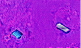
The gold standard of pseudogout diagnosis is presence of CPPD crystals in the fluid from a joint. These crystals are rhomboid-shaped structures that look either blue or red- depending on their orientation- under a polarized microscope. To find these crystals, a sample of the fluid is removed from the joint with a needle and looked at under the microscope.
How is pseudogout treated?
The goal in treatment of psedogout is to relieve the symptoms of pain and swelling.
Medications:
Non-steroidal anti-inflammatory drugs- example: ibuprofen (advil), indomethacin (Indocin), Naproxen (Naprosyn), etc.

Colchicine (Colcrys)

Steroids – example: prednisone

Pain medications like vicodin

Cold packs
Injections:
Aspiration of the fluid in the joint can help decrease the pressure and crystals that are in the joint. An injection of steroids into the joint can relieve the pain and inflammation.
Prevention of future attacks:
If risk factors are identified during the workup, those are treated to prevent future flares.

Daily colchicine (colcrys) can be used in people who experience frequent attacks of pseudogout.
Reference:
UpToDate
eMedicine http://emedicine.medscape.com/article/1268019-overview
Mayo clinic http://www.mayoclinic.com/health/pseudogout/DS00717